43 image of microscope with label
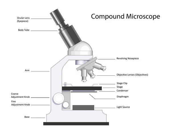
Microscope Labeling - The Biology Corner The google slides shown below have the same microscope image with the labels for students to copy. I often spend the first day walking students through the steps and having them look at a single slide as we do the steps. Students are often very enthusiastic about using microscopes and will try to start with the high power objective. Parts of a Compound Microscope - Labeled (with diagrams) It is used to carry the microscope and at the same time connect the base of the microscope to the head. (1, 2, 3, and 4) Image 3: A compound microscope with a corresponding label of the different parts.
A new holographic microscope allows scientists to see through … 16/09/2022 · It is said that the new microscope can "see through" the intact skull, and is capable of high-resolution 3D imaging of the neural network within a living mouse brain without removing the skull.

Image of microscope with label
Compound Microscope - Diagram (Parts labelled), Principle and Uses What are the 13 parts of a microscope? 1. Eyepiece 2. Eyepiece Tube 3. Objective Lens 4. Stage 5. Stage Clips 6. Nosepiece 7. Fine and Coarse Focus knobs 8. Illuminator 9. Aperture 10. Iris Diaphragm 11. Condenser 12. Condenser Focus Knob 13. The Rack stop Q 5. What are the 11 parts of a compound microscope? Compound Microscope Parts - Labeled Diagram and their Functions The eyepiece (or ocular lens) is the lens part at the top of a microscope that the viewer looks through. The standard eyepiece has a magnification of 10x. You may exchange with an optional eyepiece ranging from 5x - 30x. [In this figure] The structure inside an eyepiece. The current design of the eyepiece is no longer a single convex lens. A new holographic microscope allows scientists to see through … 16/09/2022 · Institute for Basic Science. (2022, September 16). A new holographic microscope allows scientists to see through the skull and image the brain: The new label-free deep-tissue imaging with the wave ...
Image of microscope with label. Microscope Parts and Functions First, the purpose of a microscope is to magnify a small object or to magnify the fine details of a larger object in order to examine minute specimens that cannot be seen by the naked eye. Here are the important compound microscope parts... Eyepiece: The lens the viewer looks through to see the specimen. › iet › microscopeVirtual Microscope - NCBioNetwork.org Lesson Description BioNetwork’s Virtual Microscope is the first fully interactive 3D scope - it’s a great practice tool to prepare you for working in a science lab. Explore topics on usage, care, terminology and then interact with a fully functional, virtual microscope. › releases › 2022A new holographic microscope allows scientists to see through ... Sep 16, 2022 · Institute for Basic Science. (2022, September 16). A new holographic microscope allows scientists to see through the skull and image the brain: The new label-free deep-tissue imaging with the wave ... › products › microscopeMicroscope Imaging Software | Products | Leica Microsystems Aug 23, 2021 · Microscope and digital camera configuration and control in a fully integrated manner. Basic annotation tools allow image and calibration data to be added to images. Auto and manual exposure adjustments allow optimized imaging conditions. A thumbnail gallery of acquired images, which can be reviewed quickly and easily.
Microscope Labeling Game - PurposeGames.com About this Quiz. This is an online quiz called Microscope Labeling Game. There is a printable worksheet available for download here so you can take the quiz with pen and paper.. This quiz has tags. Click on the tags below to find other quizzes on the same subject. Compound Microscope Parts, Functions, and Labeled Diagram Compound Microscope Definitions for Labels. Eyepiece (ocular lens) with or without Pointer: The part that is looked through at the top of the compound microscope. Eyepieces typically have a magnification between 5x & 30x. Monocular or Binocular Head: Structural support that holds & connects the eyepieces to the objective lenses. Parts of Stereo Microscope (Dissecting microscope) – labeled … Unlike a compound microscope that offers a flat image, stereo microscopes give the viewer a 3-dimensional image that you can see the texture of a larger specimen. [In this image] Examples of Stereo & Dissecting microscopes. Major microscope brands (Zeiss, Olympus, Nikon, Amscope, Omano, Leica …) all produce stereomicroscopes. › game › microscope-labelingMicroscope Labeling Game - PurposeGames.com This is an online quiz called Microscope Labeling Game. There is a printable worksheet available for download here so you can take the quiz with pen and paper. This quiz has tags. Click on the tags below to find other quizzes on the same subject.
Parts of a microscope with functions and labeled diagram - Microbe Notes Q. List down the 18 parts of a Microscope. 1. Ocular Lens (Eye Piece) 2. Diopter Adjustment 3. Head 4. Nose Piece 5. Objective Lens 6. Arm (Carrying Handle) 7. Mechanical Stage 8. Stage Clip 9. Aperture 10. Diaphragm 11. Condenser 12. Coarse Adjustment 13. Fine Adjustment 14. Illuminator (Light Source) 15. Stage Controls 16. Base 17. 473,906 Microscope Images, Stock Photos & Vectors | Shutterstock 473,906 microscope stock photos, vectors, and illustrations are available royalty-free. See microscope stock video clips Image type Orientation Sort by Popular Science College and University Healthcare and Medical Jobs/Professions Biology microscope laboratory scientist medicine research Next of 4,740 Microscope slide - Wikipedia A microscope slide is a thin flat piece of glass, typically 75 by 26 mm (3 by 1 inches) and about 1 mm thick, used to hold objects for examination under a microscope.Typically the object is mounted (secured) on the slide, and then both are inserted together in the microscope for viewing. This arrangement allows several slide-mounted objects to be quickly inserted and … Simple Microscope - Diagram (Parts labelled), Principle, Formula and Uses The working principle of a simple microscope is that when a lens is held close to the eye, a virtual, magnified and erect image of a specimen is formed at the least possible distance from which a human eye can discern objects clearly. Magnification formula The magnification power of a simple microscope is expressed as: M = 1 + D/F Where
Microscope Labeling Activity - SMART Board Activity - Interactive Review
Simple Microscope - Parts, Functions, Diagram and Labelling Picture 1: The image above is a stereo microscope. Image source: made-in-china.com Picture 2: The image above is a confocal microscope. Image source:thorlabs.com Picture 3: The image above is parts of scanning electron microscope. Image source:britannica.com Picture 4: The picture is a transmission electron microscope. Image source: ysjournal.com
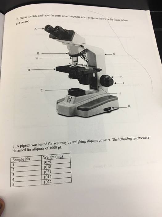
Solved Identify and label the parts of a compound microscope ...
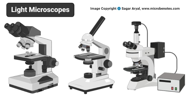
22 Parts Of a Microscope With Their Function And Labeled Diagram A light microscope is a type of microscope that commonly uses visible light and a system of lenses to generate magnified images of small objects whereas electron microscope is a microscope that uses a beam of accelerated electrons as a source of illumination. It is a special type of microscope with a high resolution of images.

Label the light microscope | Teaching Resources
Microscope Types (with labeled diagrams) and Functions The working principle of a simple microscope is that when a lens is held close to the eye, a virtual, magnified and erect image of a specimen is formed at the least possible distance from which a human eye can discern objects clearly. Simple microscope labeled diagram Simple microscope functions It is used in industrial applications like:
Microscope Labeling
BX63 | Automated Fluorescence Microscope | Olympus LS Fully motorized and easy to use, our automated, upright fluorescence microscope enables fast, efficient image capture. Learn more.
Solved Nikon Parts of the compound microscope Write the ...
18,701 Microscope drawing Images, Stock Photos & Vectors - Shutterstock Microscope drawing royalty-free images 18,701 microscope drawing stock photos, vectors, and illustrations are available royalty-free. See microscope drawing stock video clips Image type Orientation Color People Artists Sort by Popular Science Abstract Designs and Shapes College and University Art Styles Printing, Typography, and Calligraphy
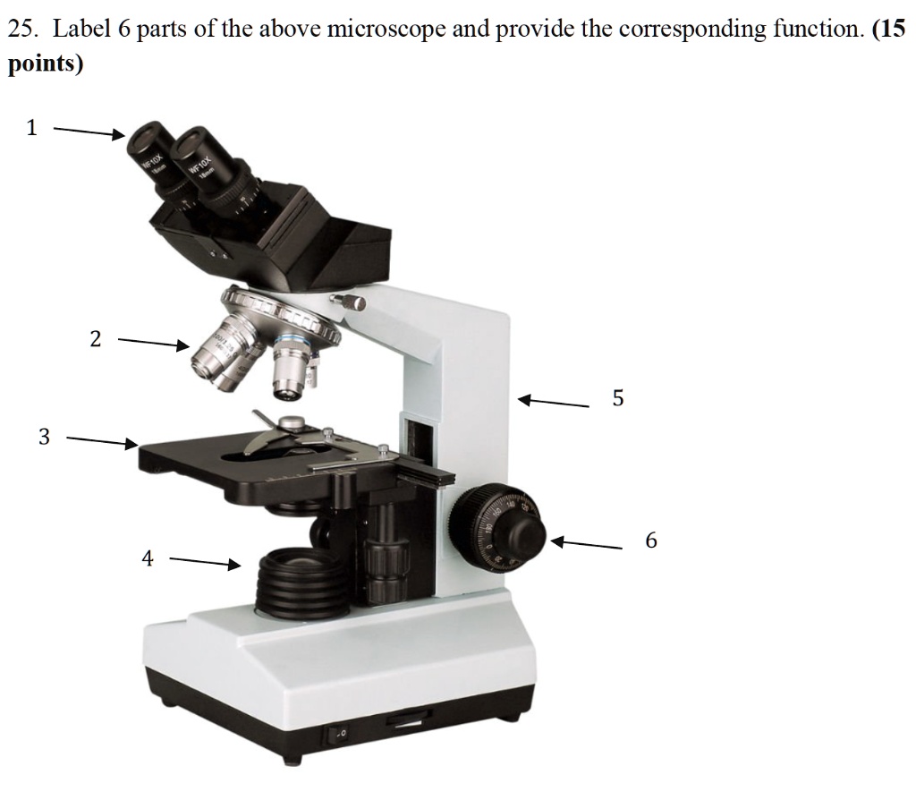
SOLVED: 25. Label 6 parts of the above microscope and provide ...
Microscope picture label Flashcards | Quizlet Start studying Microscope picture label. Learn vocabulary, terms, and more with flashcards, games, and other study tools.

Label the Parts of the Microscope - Brainly.ph
400+ Free Microscope & Bacteria Images - Pixabay 400+ Free Microscope & Bacteria Images - Pixabay 412 Free images of Microscope Related Images: bacteria laboratory science scientist research biology lab virus microscopic Find your perfect microscope image. Free pictures to download and use in your next project.

Learning Task 3: Label Me!Label The Parts of the Microscope ...
A Study of the Microscope and its Functions With a Labeled Diagram ... May 21, 2019 - To better understand the structure and function of a microscope, we need to take a look at the labeled microscope diagrams of the compound and electron microscope. These diagrams clearly explain the functioning of the microscopes along with their respective parts.

Simple Microscope - Diagram (Parts labelled), Principle ...
Microscope Parts, Function, & Labeled Diagram - slidingmotion Diaphragm. The diaphragm is also called as iris. This iris situates below the stage of the microscope. The function of the diaphragm is to control the amount of light that focuses on the specimen. This diaphragm can adjust the amount of light and intensity of light that falls on the specimen. In some standard and high-quality microscopes, this ...
4,847 Microscope labeled Images, Stock Photos & Vectors ...
Microscope With Labels clip art - Pinterest Jul 3, 2012 - Download Clker's Microscope With Labels clip art and related images now. Multiple sizes and related images are all free on Clker.com.

Compound Microscope- Definition, Labeled Diagram, Principle ...
Parts of the Microscope with Labeling (also Free Printouts) Microscopes are specially created to magnify the image of the subject being studied. This exercise is created to be used in homes and schools. the microscope layout, including the blank and answered versions are available as pdf downloads. Click to Download : Label the Parts of the Microscope (A4) PDF print version.

Biology label part of microscope
Label the microscope — Science Learning Hub Label the microscope Interactive Add to collection Use this interactive to identify and label the main parts of a microscope. Drag and drop the text labels onto the microscope diagram. eye piece lens diaphragm or iris coarse focus adjustment stage base fine focus adjustment light source high-power objective Download Exercise Tweet

Compound Microscope – Diagram (Parts labelled), Principle and ...
phys.org › news › 2022-09-holographic-microscopeA new holographic microscope allows scientists to see through ... Sep 16, 2022 · It is said that the new microscope can "see through" the intact skull, and is capable of high-resolution 3D imaging of the neural network within a living mouse brain without removing the skull.

What is a Compound Microscope? | Microscope World Blog
www1.udel.edu › biology › ketchamMicroscopy Pre-lab Activities - University of Delaware Microscope controls: turn knobs (click and hold on upper or lower portion of knob) throw switches (click and drag) turn dials (click and drag) move levers (click and drag) changes lenses (click and drag on objective housing) select a specimen (click on a slide)

Photo Compound microscope with labels Image #3850568
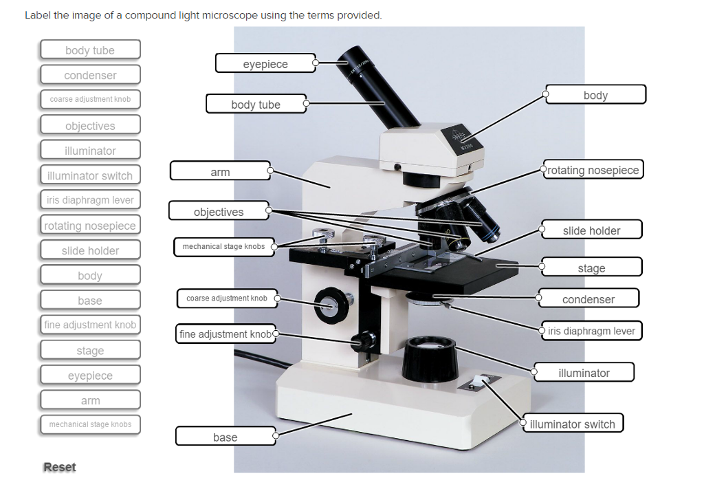
Solved Label the image of a compound light microscope using - Chegg Expert Answer. 100% (17 ratings) Transcribed image text: Label the image of a compound light microscope using the terms provided.
Label a microscope - Teaching resources
Microscope Labeled Pictures, Images and Stock Photos View microscope labeled videos Browse 49 microscope labeled stock photos and images available, or start a new search to explore more stock photos and images. Newest results Fluorescent Imaging immunofluorescence of cancer cells growing... Microscope diagram vector illustration. Labeled zoom instrument... Microscope diagram vector illustration.

Microscope - Label - Part 2 Diagram | Quizlet
Label the Model Human Cell Quiz - PurposeGames.com This online quiz is called Label the Model Human Cell #biology #anatomy. This online quiz is called Label the Model Human Cell #biology #anatomy . English en. Login. Login Register Free Help; Start; Explore. Games; Playlists; Tournaments; Tags; The Wall; Badges; Leaderboard; Create. Create a Quiz; Create a Group; Create a Playlist; Groups. Overview; Create a Group; …

This is a common compound microscope. What the labelling D ...
interestingengineering.com › innovation › novelA novel holographic microscope could image mouse brain ... Sep 19, 2022 · Here, we introduce a label-free deep-tissue imaging technique termed dimensionality reduction adaptive-optical microscopy (DReAM) to selectively attenuate multiple scattering.

Compound Microscope Parts – Labeled Diagram and their ...
Virtual Microscope - NCBioNetwork.org Lesson Description BioNetwork’s Virtual Microscope is the first fully interactive 3D scope - it’s a great practice tool to prepare you for working in a science lab. Explore topics on usage, care, terminology and then interact with a fully functional, virtual microscope. When you are ready, challenge your knowledge in the testing section to see what you have learned.

Microscope Terms Glossary | Earth science lessons, Biology ...
4847 Microscope labeled Images, Stock Photos & Vectors Find Microscope labeled stock images in HD and millions of other royalty-free stock photos, illustrations and vectors in the Shutterstock collection.

Compound Microscope Parts, Diagram Definition, Application ...
Microscope Imaging Software | Products | Leica Microsystems 23/08/2021 · Microscope and digital camera configuration and control in a fully integrated manner. Basic annotation tools allow image and calibration data to be added to images. Auto and manual exposure adjustments allow optimized imaging conditions. A thumbnail gallery of acquired images, which can be reviewed quickly and easily.
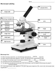
Microscope Labeling - Name_ Microscope Labeling occular lens ...
Electron Microscope Principle, Uses, Types and Images (Labeled Diagram ... The cost of a new electron microscope ranges between $80,000 to $10,000,000 and above depending on the customizations, configurations, resolution, components, and brand value. The type of electron microscope also decides the price of the microscope because of the various uses the microscope has and also on the components used in them.

A Study of the Microscope and its Functions With a Labeled ...
Microscope With Labels Clip Art at Clker.com Download Clker's Microscope With Labels clip art and related images now. Multiple sizes and related images are all free on Clker.com.

Label the microscope — Science Learning Hub
Compound Microscope- Definition, Labeled Diagram, Principle, Parts, Uses The optical microscope often referred to as the light microscope, is a type of microscope that uses visible light and a system of lenses to magnify images of small subjects. The term "compound" in compound microscopes refers to the microscope having more than one lens. Devised with a system of combination of lenses, a compound microscope ...

This is a common compound microscope. Label its parts from A ...
A novel holographic microscope could image mouse brain … 19/09/2022 · Researchers from various universities are now viewing the mouse brain through the skull thanks to a new holographic microscope. The device can provide high-resolution 3D imaging of the neural network.
Microscope slide Vector Art Stock Images | Depositphotos
Labeling the Parts of the Microscope | Microscope World Resources Labeling the Parts of the Microscope This activity has been designed for use in homes and schools. Each microscope layout (both blank and the version with answers) are available as PDF downloads. You can view a more in-depth review of each part of the microscope here. Download the Label the Parts of the Microscope PDF printable version here.
Guide to using a microscope - Home
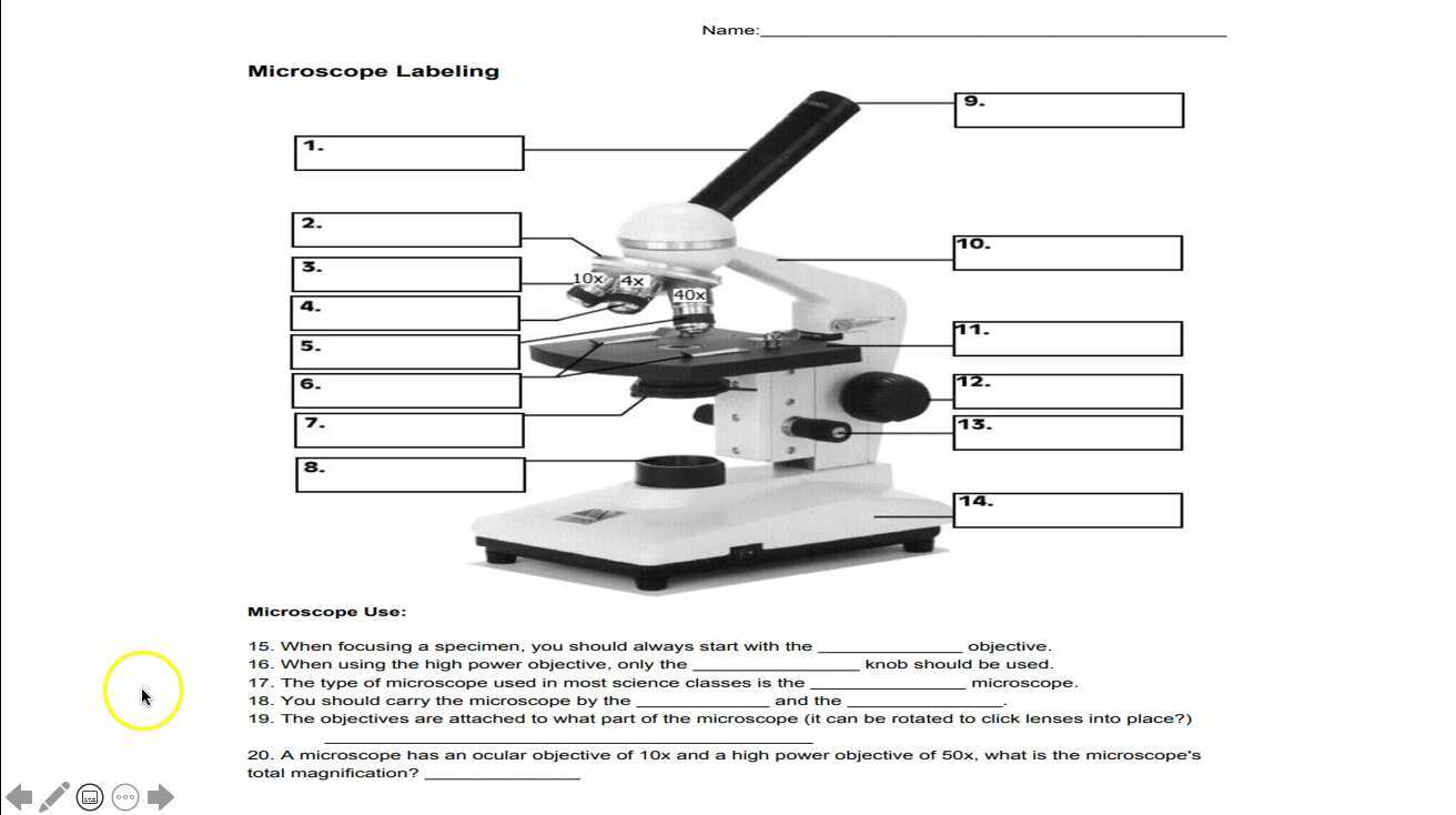
Microscope Labeling - The Biology Corner Students label the parts of the microscope in this photo of a basic laboratory light microscope. Can be used for practice or as a quiz. ... Microscope Labeling . Microscope Use: 15. When focusing a specimen, you should always start with the _____ objective. 16. When using the high power objective, only the _____ knob should be used. 17. The ...

Simple Microscope - Diagram (Parts labelled), Principle ...
Binocular Microscope Pictures, Images and Stock Photos Browse 814 binocular microscope stock photos and images available, or search for microtome or histology to find more great stock photos and pictures. Young Asian researcher using modern microscope in laboratory. Young Asian researcher using a modern microscope in a laboratory.
Transmitted light microscope B3 Professional series B3-220ASC ...
Compound Microscope Labeled Diagram | Quizlet Part that supports the microscope. Stage. Supports the slide or specimen. Coarse adjustment Knob. sed to focus when using the low power objective lenses. Fine Adjustment Knob. Used to focus the image on high power to view image in more detail. Revolving nose piece. The revolving piece on which the lenses are attached.

Optical microscope, microscope, angle, simple, label png ...
Microscope, Microscope Parts, Labeled Diagram, and Functions Microscope, Microscope Parts, Labeled Diagram, and Functions What is Microscope? A microscope is a laboratory instrument used to examine objects that are too small to be seen by the naked eye. It is derived from Ancient Greek words and composed of mikrós, "small" and skopeîn,"to look" or "see".
Light Microscope- Definition, Principle, Types, Parts ...
Parts of a Simple Microscope - Labeled (with diagrams) A simple microscope is a very first type of microscope ever created. It consists of simple parts and performs simple functions. Although there are now many advanced microscope types, some applications may still demand the use of a simple microscope. In this article, we are going to discuss the parts and functions of a simple microscope.

Microscope labeled diagram
Microscope labeled diagram - SlideShare The Microscope Image courtesy of: Microscopehelp.com Basic rules to using the microscope 1. You should always carry a microscope with two hands, ...

label the parts of the compound microscope - Brainly.ph
A new holographic microscope allows scientists to see through … 16/09/2022 · Institute for Basic Science. (2022, September 16). A new holographic microscope allows scientists to see through the skull and image the brain: The new label-free deep-tissue imaging with the wave ...
Lab - Microscope: MAH-Summer 2019-Anatomy and Physiology I
Compound Microscope Parts - Labeled Diagram and their Functions The eyepiece (or ocular lens) is the lens part at the top of a microscope that the viewer looks through. The standard eyepiece has a magnification of 10x. You may exchange with an optional eyepiece ranging from 5x - 30x. [In this figure] The structure inside an eyepiece. The current design of the eyepiece is no longer a single convex lens.

Compound Microscope Labeled Diagram | Quizlet
Compound Microscope - Diagram (Parts labelled), Principle and Uses What are the 13 parts of a microscope? 1. Eyepiece 2. Eyepiece Tube 3. Objective Lens 4. Stage 5. Stage Clips 6. Nosepiece 7. Fine and Coarse Focus knobs 8. Illuminator 9. Aperture 10. Iris Diaphragm 11. Condenser 12. Condenser Focus Knob 13. The Rack stop Q 5. What are the 11 parts of a compound microscope?

Microscope Parts and Functions

Parts of a microscope with functions and labeled diagram
Solved Label the image of a compound light microscope using ...

Label the Microscope Diagram | Download Scientific Diagram

Microscopy and Its Types - BIOLOGY EASE

The Microscope- compound microscope diagram - Major Science ...

Microscope With Labels clip art | Microscope parts ...

Virtually Labeling a Microscope by Grace Voit | Teachers Pay ...


Komentar
Posting Komentar